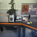
2ая школа для молодых ученых по современным методам флуоресцентной микроскопии (ADFLIM)

Research Resource center
(ext. number 6640)
+7(812)363-60-39

 08.02.2023
Компания БМТ совместно с компанией Sartorius приглашает вас на обучающий семинар «Академия дозирования» для специалистов лабораторий.
 15.05.2021
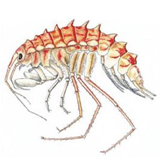
18 мая Полина Борисовна Дроздова сделает доклад на тему: "Свет, цвет и байкальские амфиподы".
 12.05.2021
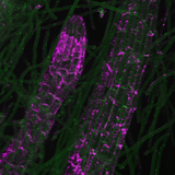
14 мая в 15:00 состоится семинар по теме «Применение лектинов для выявления растительных гликанов методом флуоресцентной in situ гибридизации».
 28.04.2021
4 мая 2021, в 14:00. Лекция нобелевского лауреата Штефана Хелля
 07.09.2020
с 29 сентября по 2 октября, международная конференция «Towards optical and multimodality translational imaging»
• 3D (xyz) и 4D (xyzt) scanning of various biological objects,including resonance scanning of fast moving entity (whole microorganisms and live cell organelles), fluorochrome emission andautofluorescence scanning (λ-scanning), FLIP (fluorescence loss in photobleaching), FRAP (fluorescence recovery after photobleaching), FRET (fluorescence resonance energy transfer);
• analysis of the structure and macromolecular composition of biological objects by using wide spectrum applications of PCR (polymerase chain reaction) and FISH (fluorescence in situ hybridization), such as CGH (comparative genomic hybridization), PRINS (primed in situ synthesis), RT (reverse in situ transcription), WMISH (whole mount in situ hybridization), and immunocytochemistry;
• cell microsurgery, micromanipulations and microinjections of the cells and tissues;
• TEM ultrastructural studies and visualization of macromolecules;
• bioimaging: 3D-reconstruction, deconvolution and statistical analysis.
